Placenta Characterization
The placenta forms the critical interface between the fetus and the mother, and is responsible to provide a safe and protective milieu for fetal development. Placental dysfunction plays a central role in the development of pre-eclampsia, fetal growth restriction, and pre-term labor and may be responsible for a significant proportion of maternal and perinatal mortality and morbidity. Placental disorders involve both macro- and microstructural changes of the tissue. Quantitative ultrasound (QUS), with the ability to characterize tissue microstructure, is a suitable candidate for developing an effective screening tool. Among the QUS parameters, attenuation coefficient estimate (ACE) and shear wave speed could be potential biomarkers to detect complications in pregnancy. We have explored the feasibility of characterizing placental tissue using both the shear wave speed and ACE, using radio frequency (RF) data collected from a shear wave vibro-elastography (SWAVE) study [1]. Placenta specimens were subject to an external excitation that generated a shear wave within them and an ultrasound scanner was used to measure the shear wave speed. The ultrasound radio frequency (RF) data acquired while applying the mechanical excitation contained information from both the shear wave and the longitudinal wave. These two entities conveyed qualitatively different information due to differences in wavelength. report the baseline attenuation coefficient based on a large cohort of clinically normal placentas (n=59) [2, 3].
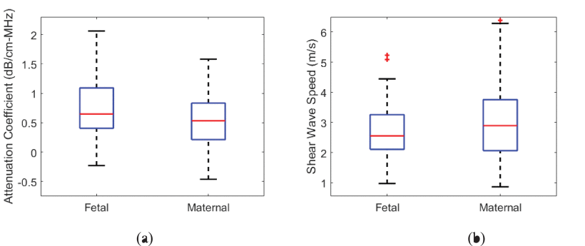
Figure: Baseline values for ACE and shear wave speed based on 59 clinically normal full-term placentas. Box and whisker representation of (a) ACE, and (b) shear wave speed for region-of-interest near the fetal surface and near the maternal surface.
We observed cases where the ACE map and cs map show a similar pattern indicating a common influence by the underlying structure. The shear wave speed and ACE values appeared to be spatially correlated. However, the correlation pattern varied for different cases. We hypothesize that the underlying structure of the placenta is heterogeneous, so we expect different correlation patterns between ACE and shear wave speed for different underlying structures.
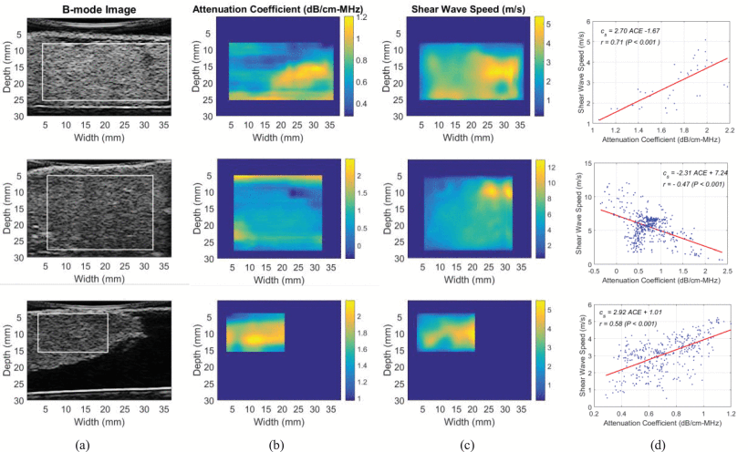
Figure: Example images of attenuation coefficient map and shear wave speed map. (a) Ultrasound B-mode image with region of interest
(highlighted in white) used for ACE and shear wave speed measurement, (b)
attenuation coefficient map, (c) shear wave speed map, and (d) correlation between attenuation coefficient and shear wave speed. <\h5>
We are currently conducting SWAVE 2.0 to acquire data from 20 normal and 40 abnormal placentas. After the study completion, we will analyze the data and investigate the potential of the simultaneously measured QUS parameters to distinguish between normal and pathologic placentas.
1. Abeysekera, Jeffrey M., et al. "SWAVE imaging of placental elasticity and viscosity: proof of concept." Ultrasound in Medicine and Biology 43.6 (2017): 1112-1124.
2.Deeba, Farah, et al. "Multiparametric QUS Analysis for Placental Tissue Characterization." 2018 40th Annual International Conference of the IEEE Engineering in Medicine and Biology Society (EMBC). IEEE, 2018.
3. Deeba, Farah, et al. "Attenuation coefficient estimation of normal placentas." Ultrasound in medicine & biology 45.5 (2019): 1081-1093.